Tiedosto:Fundus of patient with retinitis pigmentosa, mid stage.jpg
Tämän esikatselun koko: 699 × 599 kuvapistettä. Muut resoluutiot: 280 × 240 kuvapistettä | 560 × 480 kuvapistettä | 871 × 747 kuvapistettä.
Alkuperäinen tiedosto (871 × 747 kuvapistettä, 107 KiB, MIME-tyyppi: image/jpeg)
| Tämä tiedosto on tiedostotietokanta Wikimedia Commonsista. Tiedot kuvaussivulta näkyvät alla. |  |
Tiedoston kuvaussivu Commonsissa |
| Kuvaus |
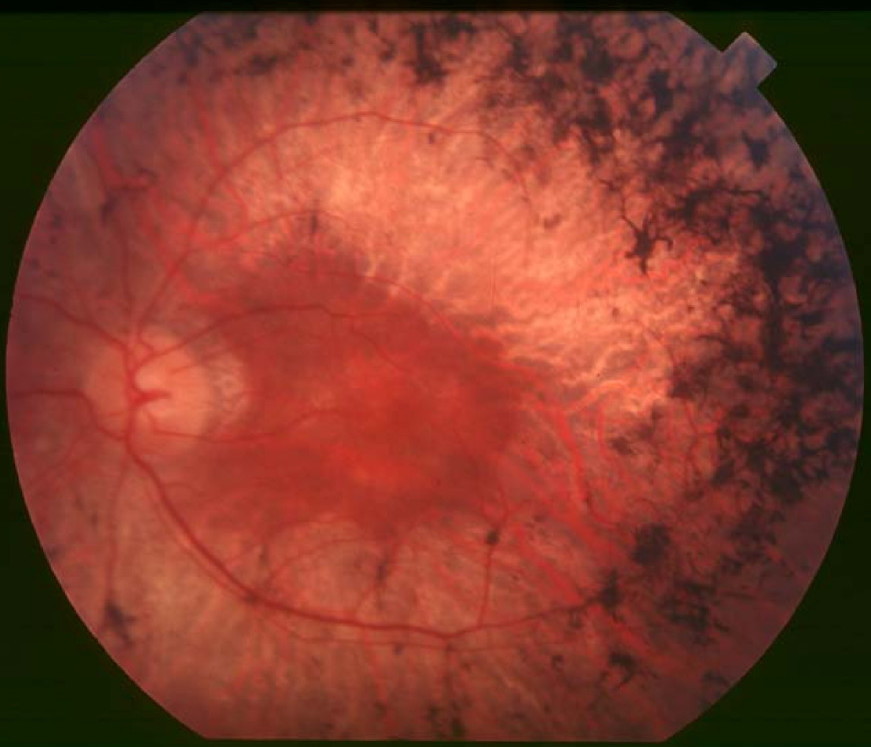
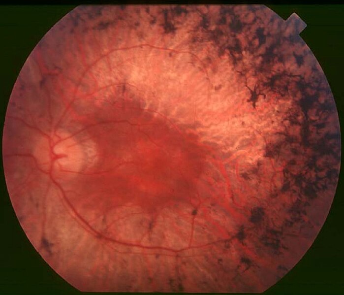
English: Figure 2. Fundus of patient with retinitis pigmentosa, mid stage (Bone spicule-shaped pigment deposits are present in the mid periphery along with retinal atrophy, while the macula is preserved although with a peripheral ring of depigmentation. Retinal vessels are attenuated.) Hamel Orphanet Journal of Rare Diseases 2006 1:40 doi:10.1186/1750-1172-1-40 |
| Päiväys | |
| Lähde | Retinitis pigmentosa by Christian Hamel |
| Tekijä | Christian Hamel |
| Käyttöoikeus (Tämän tiedoston uudelleenkäyttö) |
© 2006 Hamel; licensee BioMed Central Ltd. This is an Open Access article distributed under the terms of the Creative Commons Attribution License (https://creativecommons.org/licenses/by/2.0), which permits unrestricted use, distribution, and reproduction in any medium, provided the original work is properly cited. |
Tämä tiedosto on lisensoitu Creative Commons Nimeä 2.0 Yleinen -lisenssillä.
- Voit:
- jakaa – kopioida, levittää ja esittää teosta
- remiksata – valmistaa muutettuja teoksia
- Seuraavilla ehdoilla:
- nimeäminen – Sinun on mainittava lähde asianmukaisesti, tarjottava linkki lisenssiin sekä merkittävä, mikäli olet tehnyt muutoksia. Voit tehdä yllä olevan millä tahansa kohtuullisella tavalla, mutta et siten, että annat ymmärtää lisenssinantajan suosittelevan sinua tai teoksen käyttöäsi.
Tiedoston historia
Päiväystä napsauttamalla näet, millainen tiedosto oli kyseisellä hetkellä.
| Päiväys | Pienoiskuva | Koko | Käyttäjä | Kommentti | |
|---|---|---|---|---|---|
| nykyinen | 2. joulukuuta 2017 kello 13.17 | | 871 × 747 (107 KiB) | Doc James | Cropped 27 % horizontally and 7 % vertically using CropTool with precise mode. |
| 22. syyskuuta 2009 kello 16.52 |  | 1 200 × 799 (126 KiB) | CopperKettle | {{Information |Description={{en|1=Figure 2. Fundus of patient with retinitis pigmentosa, mid stage (Bone spicule-shaped pigment deposits are present in the mid periphery along with retinal atrophy, while the macula is preserved although with a peripheral |
Tiedoston käyttö
Seuraava sivu käyttää tätä tiedostoa:
Tiedoston järjestelmänlaajuinen käyttö
Seuraavat muut wikit käyttävät tätä tiedostoa:
- Käyttö kohteessa ar.wikipedia.org
- Käyttö kohteessa bs.wikipedia.org
- Käyttö kohteessa ca.wikipedia.org
- Käyttö kohteessa da.wikipedia.org
- Käyttö kohteessa en.wikipedia.org
- Käyttö kohteessa en.wikiversity.org
- Käyttö kohteessa es.wikipedia.org
- Käyttö kohteessa eu.wikipedia.org
- Käyttö kohteessa fa.wikipedia.org
- Käyttö kohteessa fr.wikipedia.org
- Käyttö kohteessa he.wikipedia.org
- Käyttö kohteessa hy.wikipedia.org
- Käyttö kohteessa it.wikipedia.org
- Käyttö kohteessa ko.wikipedia.org
- Käyttö kohteessa la.wikipedia.org
- Käyttö kohteessa or.wikipedia.org
- Käyttö kohteessa outreach.wikimedia.org
- Käyttö kohteessa pl.wikipedia.org
- Käyttö kohteessa pt.wikipedia.org
- Käyttö kohteessa ru.wikipedia.org
- Käyttö kohteessa sl.wikipedia.org
- Käyttö kohteessa sr.wikipedia.org
- Käyttö kohteessa sv.wikipedia.org
- Käyttö kohteessa th.wikipedia.org
- Käyttö kohteessa tr.wikipedia.org
- Käyttö kohteessa tt.wikipedia.org
- Käyttö kohteessa uk.wikipedia.org
- Käyttö kohteessa vi.wikipedia.org
Näytä lisää tämän tiedoston järjestelmänlaajuista käyttöä.